- The endoplasmic reticulum (ER) is an irregular network of branching and fusing membranous tubules, around 40 to 70 nm in diameter, and many flattened sacs called cisternae (s., cisterna).
- The nature of the ER varies with the functional and physiological status of the cell.
- In cells synthesizing a great deal of protein to be secreted, a large part of the ER is studded on its outer surface with ribosomes and is called rough endoplasmic reticulum .
- Other cells, such as those producing large quantities of lipids, have ER that lacks ribosomes. This is smooth endoplasmic reticu lum (SER).
- The endoplasmic reticulum has many important func tions.
- Not only does it transport proteins, lipids, and other materials through the cell, it is also involved in the synthesis of many of the materials it transports.
- Lipids and proteins are synthesized by ER-associated enzymes and ribosomes.
- Poly peptide chains synthesized on RER-bound ribosomes may be inserted either into the ER membrane or into its lumen for transport elsewhere.
- The ER is also a major site of cell mem brane synthesis.
Sunday, August 22, 2021
Endoplasmic Reticulum

immunohematology,Blood group genteics,Blood Group Antibodies,Autoantibodies,Main Blood group system,
Introduction:
• Each species of animals , including humans has certain antigens on the surface of the red cells which are unique for that species.
• These are known as isoantigens. Similarly certain antigens , the alloantigens are common to some, but not all members of that particular species.
• Blood group serology involves the detection of these antigens & their antibodies.
Blood group genteics:
• All the antigenic substances present on RBC of indiviual are inherited. The unit of inheritance is a gene & each antigen is controlled by gene for that antigen.
• The position of each gene on a chromosome is called its locus.
• Some terminology:
• Allele or allelomorph: Two or more genes responsible for same chracteristics occupying the same position on the chromosomes.
• Homozygous: A person who has inherited same allelic genes from both parents eg: having both blue eyed colour genes.
• Hetrogygous: A person who has inherited different allelic genes for trait. eg: having a blue & brown eye colour genes.
Dominat gene: Certain alleles are stronger than other alleles. eg: brown eyes genes mask the presence of blue eye genes, & are therefore dominant over blue eye genes. A dominat gene expresses itself in both homozygous & hetrozygous states.
• Recessive gene: They are expressed only when dominant allele is absent eg: in Homogygous state. Colour of eye is blue ,if both alleles are for blue eyes.
• Co-dominat gene: In this ,various alleles for the same character are equally dominant eg: alleles for a particular blood group system.
• Genotype: The genetic make up of an individual or cell is called its genotype.
• Phenotype: It is the observable characteristics of individual or a cell eg: an individual of group Bo genotype would be expressed as group B because O gene is rcessive.
Blood Group Antigens:
• The blood group antigens include substances on the RBC & on other consitutents such as leucocytes, platelets & plasma.
• The blood consitutent antigens which are governed by single gene or a group of genes are inherited as groups.
• Blood groups controlled by set of genes are called a blood group system.
• Most blood group antigens follow the law of inheritance. Chemically blood group antigens are usually glycoproteins, lipoproteins or glycolipids in nature.

Blood Group Antibodies:
• Specific blood antibodies develop in response to antigenic stimulation by particular blood group antigen.
• The level of antibody activity depends on the immunogenicity of corresponding antigen.
• Alloantidodies:
• Are antibodies which are present in some members of species but not all. eg: anti-A antibodies are present in all
humans beigns belonging to blood B & O.
• Due to the presence of alloantibodies, it is necessary to select a blood donor who is negative for corresponding antigen,i.e if recipient has anti-A antibodies, he cannot recieve blood which contain group A antigen.
• They are of three type:
1. Naturally occurring: The antigenic stimulus is unknown. ABO antibodies belong to this type.
2. As a result of immunization through transfusion, some red cell antigens which are absent in the recipent may be introduced & induce antibody production.
3. Induced by exposure to foetal erythrocytes either during pregneancy or during delivery. These antibodies are similar to those produced by blood transfusion. Development of Rh antibodies in a Rh negative mother by a Rh positive foetus is good example.

Autoantibodies:
• An autoantibody is the antibody which is induced by an antigen in same individual. It also reacts with same antigen if it present in other individual.
• In some cases the reaction of antigen & its autoantibody may not show any demonstratable clinical symptoms, whereas sometimes such a reaction may lead to haemolytic anaemia, leucopenia or thrombocytopenia.
• It can be divided into two general catagories depending on their optimum reacting Temperature.
1. Warm autoantibodies: These consitutue about 85% of autoantibodies. Their optimal temperature is 370C. They are generally IgG in nature & can be detected with direct antibody test.
2. Cold auto antibodies: About 15% are of this type. they are generally IgM in nature & agglutinate RBC strongly at 40C weakly at 240C & not at all 370C. Most of are present in normal individual, very few are associated with diseases. It is important to be aware of cold antibodies, when the patients body temperature needs to be lowered for procedures such as cardiopulmonary bypass.
: Main Blood group system:
• Approximately 600 blood group antigens have been describes so far.
• The konwoledge of blood group antigens is important during blood transfusion.
• Among these ABO system is most important blood group system in blood transfusion & in organ transplantation because of two unique features:
1. Strongly reactive antibodies are present in sera of individual who lack the corresponding antigens:
2. A & B antigens are present on many tissues cells in addition to red cells.
• The ABO system consist of four blood groups or phenotypes : A, B, AB & O. The two antigen A & B are responsible for these four groups.
• If A antigen is present on red cell individual is said to belong to A group ,If B antigen is present on red cell individual is said to belong to B group, & If AB antigen is present on red cell individual is said to belong to AB group.
- While O groups have neither A or B antigen on their red cell.
• Three allelic genes A, B & O,can be inherited in ABO system. The following combination of alleles is
possible: AA, AO, AB, BB, BO, OO resulting in A, A, AB, B, B & O group individuals respectively.
• This so because A & B genes are dominat & O gene is recessive.

Law of Inheritance of ABO Groups:
• According to Bernstein's theory two laws of inheritance have been proved.
1. It is not possible that an offspring can possess antigen A, B or both unless it is inherited from one or both parents.
2. A parent of blood group AB cannot produce an offspring of group O. Similarly a parent of blood group O cannot produce AB offspring. The reason for this law is that group AB is hetrozygygous & therefore A & B genes must come from two parents.
Group O individual have antigen called H antigen on the surface of red cells , which is precusssor of A & B antigen.
Subgroups of ABO:
• Both groups A & B can be further subdivided . The most important of these subgroups are A1, A2. Both A1 & A2 cells react with anti A antiserum.
• Approximately 20% of group A & AB persons belong to subgroup A2 & 80% to A1. Several subgroups of B have been identified but they are very rare.
Rh Grouping System, rh blood type, rh blood, how to determine rh blood type
:
• The discovery of Rh system is based on the work by Landstenier & wiener in 1940 & by Levine & Stetson in 1939.
• A woman who delivered a still- born foetus was transfused with her husband's blood. Although both husband & wife belonged to O blood group, the woman experinced a severe haemolytic reaction .
• Levine & Stetson proposed a theory that woman's red cells were lacking in an antigen & this antigen was called new antigen., which child had inherited from father.
• The antigen on foetal cells stimulated the production of antibodies in the mother's blood, so when she was transfused with husband's blood, these antibodies brought about haemolytic reaction.
• In 1940, Landsteiner & Wiener inoculated red cells of Rhesus monkey into rabbits & guinea pigs. The resulting antibodies agglutinated the red cells of monkeys & also of about 85% of the population.
These 85% were called Rh (Rhesus) positive because they possessed the same antigen that were present on red cells of Rhesus monkeys.
• The rest of the population were called Rhesus negative . Thus it was found that antibodies to same antigen can cause haemolytic reaction.
• The designation Rh is derived from the use of the blood of rhesus monkeys in the basic test for determining the presence of the Rh antigen in human blood.
• The Rh blood group system was discovered in 1940 by Karl Landsteiner and A.S. Weiner. Since that time a number of distinct Rh antigens have been identified, but the first and most common one, called RhD, causes the most severe immune reaction and is the primary determinant of the Rh trait.
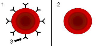
Rh Antigen:
• The Rh blood group system has two sets of nomenclatures: one developed by Ronald Fisher and R. R. Race, the other by Wiener. Both systems reflected alternative theories of inheritance. The Fisher–Race system, which is more commonly in use today, uses the CDE nomenclature.
• This system was based on the theory that a separate gene controls the product of each corresponding antigen (e.g., a "D gene" produces D antigen, and so on). However, the d gene was hypothetical, not actual.
• The Wiener system used the Rh–Hr nomenclature. This system was based on the theory that there was one gene at a single locus on each of the 2 copies of chromosome 1, each contributing to production of multiple antigens. In this theory, a gene R1 is supposed to give rise to the “blood factors” Rh0, rh′, and rh″ (corresponding to modern nomenclature of the D, C, and E antigens) and the gene r to produce hr′ and hr″ (corresponding to modern nomenclature of the c and e antigens).
• The Rh blood group system is much more complex than ABO system. More than 40 antibodies have been describe
The proteins which carry the Rh antigens are transmembrane proteins, whose structure suggest that they are ion channels.
• The main antigens are D, C, E, c and e, which are encoded by two adjacent gene loci, the RHD gene which encodes the RhD protein with the D antigen (and variants)and the RHCE gene which encodes the RhCE protein with the C, E, c and e antigens (and variants).
• There is no d antigen. Lowercase "d" indicates the absence of the D antigen (the gene is usually deleted or otherwise nonfunctional).
• Thus, there are six related blood group factors C, D, E, c, d & e and corresponding antibodies (expect anti-d).
• Like ABO antigens , the Rh factors are also inherited traits. There are six Rhesus genes C, D ,E ,c, d & e.
• Thus there are only eight possible combinations in which chromosomes can carry these genes. they are CDe, cDE, CDE,cDe, Cde, cdE, CdE & cde.
A shorthand system has been devised for easy identification of these combination.
• The three pairs of genes are carried on the same chromosome & have three closely linked loci. Every individual has loci for six Rh genes.
• The factors C, D, E, c, d, & e (expect d) are antigenic. They are capable of stimulating the production of antibodies if introduced into the body of an individual whose red cells lack them.
• However Rh antigens vary in their degree of antigenicity. The D antigen is most immunogenic of them.
• The first transfusion of Rh (D) antigen into Rh (D) negative person will stimulate the production of anti D antibodies, thus subsequent transfusion with Rh (D) antigen will result in haemolytic transfusion reaction, similar to that of Levine & Stetson's patient in 1939.
• Thus it is necessary to test the blood of both donor & the recipent for Rh D antigen before blood trnasfusion. If blood sample shows presence of Rh (D) antigen it is considered as Rh positive or show absence considered as Rh negative.

The Du antigen:
• Some D antigens react weakly with anti-D sera. This weak reactivity with anti-D sera is due to Du antigen.
• Some workers belive that the presence of this antigen can be result of some genetic variation. One theory to explain Du antigen suggest that the D antigen is made up four fractions.
• An Rh-D positive individual possesses all four , while none is present in Rh-D ne
gative person.
• Some person may lack one or more of these fractions & therefore their red cells are agglutinated by only some of the anti-D antisera.
• Such incomplete D antigen is termed as Du . It does not usually react with complete anti-D, but it react with incomplete anti-D antisera with a varying degree of reactivity.
• While typing a blood sample for Rh antigen, the antierum will not produce agglutination with Du antigen, but will produce sensitized cells i.e the red cells will be coated with antibody.
The presence of sensitisied cells can be detected by the antiglobulin test.
• If Du person is given Rh-D positive blood, he or she is likely to produce anti-D antibodies therefore he or she is considered as Rh negative recipient.
• Similarly a Du person blood, when given to Rh negative person will stimulate anti-D antibodies.
• Therefore Du blood is given only to Rh positive persons.
A Coombs test, also known as antiglobulin test (AGT) is either of two blood tests used in immunohematology. They are the direct and indirect Coombs tests. The direct Coombs test detects antibodies that are stuck to the surface of the red blood cells.
• Since these antibodies sometimes destroy red blood cells, a person can be anemic and this test can help clarify the condition. The indirect Coombs detects antibodies that are floating freely in the blood.These antibodies could act against certain red blood cells and the test can be done to diagnose reactions to a blood transfusion.
• The direct Coombs test is used to test for autoimmune hemolytic anemia—that is, a condition where the immune system breaks down red blood cells, leading to anemia.
The direct Coombs test is used to detect antibodies or complement proteins attached to the surface of red blood cells.
• To perform direct test, a blood sample is taken and the red blood cells are washed (removing the patient's own plasma and unbound antibodies from the red blood cells) and then incubated with anti-human globulin ("Coombs reagent").
• If the red cells then agglutinate, the direct Coombs test is positive, a visual indication that antibodies or complement proteins are bound to the surface of red blood cells and may be causing destruction of those cells.
• The indirect Coombs test is used in prenatal testing of pregnant women and in testing prior to a blood transfusion. The test detects antibodies against foreign red blood cells.
• In this case, serum is extracted from a blood sample taken from the patient. The serum is incubated with foreign red blood cells of known antigenicity. Finally, anti-human globulin is added. If agglutination occurs, the indirect Coombs test is positive.
• Rh antibodies:
• Unlike ABO antibodies ,there are no naturally occuring Rh antibodies. All Rh antibodies are immune antibodies ,resulting from specific antigenic stimulation. eg: transfusion, pregnancy or by injection of antigen.
• Because there are no natural Rh antibodies, antibody typing is not possible in Rh system. Therefore, all Rh typing methods depend upon antigen typing using known serum.
• Some Rh antibodies cannot be detected in saline suspension of red cells. However if a protein rich medium such as serum or albumin is used, the antibodies can agglutinate the respective red cells. such antibodies are called imcomplete or albumin active antibodies.
• Th Rh antibodies which can react even in saline solution are called complete or saline active antibodies.
• There is still another class of antibodies which can be demonstrated only by means of antihuman globulin or Coomb's reagent. Such antibodies are known as incomplete univalent antibodies
The size of antibody molecule is largely responsible for the differences in their reactivities.
• The saline active antibody molecules are of IgM type & are largest.
• The length of IgM molecule is sufficient to cause bridging of adjacent red cells in suspension. When in suspension red cells carry an electrical charge called zeta potential which causes repulsion among two adjacent red cells.
• The IgM antibody molecules extend beyond the range of zeta potential & agglutinate the red cells by binding onto their antigenic sites.
• The IgG molecules are smaller in size & cannot reach beyond the minimum distance between the cells, & are unable to agglutinate them.
• The zeta potential can be reduced by suspending the cells in high protein medium eg: patient own serum or 22% albumin solution.
• Similarly the antihuman globulin can detect incomplete univalent antibodies by bringing them together along with red cells to which they are attached.
• Rh typing Methods:
• As there are no naturally occuring Rh antibodies, Rh typing methods involve typing of red cells using known antisera against various Rh antigens.
• Commericial antisera are avaliable for C, D, E, c & e factors. since anti-d antibody is not known to be present. it is not possible to test for Rh-d factor.
• All blood sample which do not give positive reaction for Rh-D should be tested for Du antigen before reporting them as Rh-D negative.
• The method used for Rh-D typing depends upon the type of antibody used.
Method:1 Saline Method for Rh-D typing using complete Anti-D:
• A: Slide Test:
• Specimen : Whole blood or 50% red cell suspension prepared from clotted blood in patient own serum.
• Reagents: anti-D antiserum ( complete)
• Method:
1. Place a drop of antiserum on a labelled white tile.
2. Add two drops of specimen (whole blood or 50% red cell suspension).
3. Mix well & place the slide on a warm viewing box,to bring the temperature of mixture to about 370C.
4. Gently rotate the tile back & forth for a maximum of two minutes.
5. Examine microscopically for agglutination by transferring a small volume on slide with a clean pasteur pipette.
Test a positive & a negative control in the same way.
• Result:
• Agglutination: RhD positive.
• No agglutination: Rh-D negative
B: Tube Test:
• Specimen: Washed 5% red cell suspension of patient's blood.
• Reagent: Anti-D antiserum (complete)
Method:
1. In an appropriately labelled test tube add, one drop of anti-D antiserum.
2. Add one drop of 5% red cell suspension.
3. Mix well & incubate at 370C in a water bath for 30 minutes.
4. Roll the tube gently to redisperse the cells.
5. Check for agglutination microscopically. If no agglutination occurs, centrifuge at 200g for 1-2 minutes & check again.
6. Run known positive & negative controls simultaneously with test
Method 2: Albumin displacement technique for Rh typing using incomplete Anti-D:
• A 22% bovine albumin solution used in this method reduces the zeta potential (repllent electric charge) , thus bringing the red cells closer, so that IgG antibody molecules can agglutinate them.
• Specimen: 5% washed red cells suspension of patient blood.
Reagents:
• Incomplete anti-D antiserum.
• 22% bovine albumin.
Method:
1. In an appropriately labelled test tube, add one drop of incomplete anti-D & one drop of 5% red cell suspension.
2. Mix well & incubate at 370C in water bath for 60 minutes.
3. Without distrubing the cell button , run down one drop of 20% bovine albumin along the side of the tube onto cells. Do not mix.
4. Incubate further at 370C for 30 minutes.
5. Read microscopically for agglutination.
6. Run positive & negative controls simultaneously with test.
Method:3 Enzyme Techniques:
• Some antibodies aglutinate or lyse red cells which been treated with proteolytic enzymes such as papain, trypsin or bromeline.
• These enzymes remove some negatively charged molecules from red cells surface, allowing them to come closer for IgG molecules to agglutinate them.
• These proteolytic enzymes also increase agglutination by removing a part of hydration layer surrounding the red cells.
Biochemistry of H, A, & B antigen
Biosynthesis of the H, A and B antigens involves a series of enzymes (glycosyl transferases) that transfer monosaccharides.
• The resulting antigens are oligosaccharide chains, which are attached to lipids and proteins that are anchored in the red blood cell membrane. The function of the H antigen, apart from being an intermediate substrate in the synthesis of ABO blood group antigens, is not known, although it may be involved in cell adhesion.
• People who lack the H antigen do not suffer from deleterious effects, and being H-deficient is only an issue if they need a blood transfusion, because they would need blood without the H antigen present on red blood cells.
• The specificity of the H antigen is determined by the sequence of oligosaccharides. More specifically, the minimum requirement for H antigenicity is the terminal disaccharide fucose-galactose, where the fucose has an alpha(1-2)linkage. This antigen is produced by a specific fucosyl transferase that catalyzes the final step in the synthesis of the molecule.
• Depending upon a person's ABO blood type, the H antigen is converted into either the A antigen, B antigen, or both. If a person has group O blood, the H antigen remains unmodified. Therefore, the H antigen is present more in blood type O and less in blood type AB.
• Hh antigen system - diagram showing the molecular structure of the ABO(H) antigen system.
• Two regions of the genome encode two enzymes with very similar substrate specificities: the H locus (FUT1) which encodes the Fucosyl transferase and the Se locus (FUT2) that instead indirectly encodes a soluble form of the H antigen, which is found in bodily secretions. Both genes are on chromosome 19 at q.13.3. - FUT1 and FUT2 are tightly linked, being only 35 kb apart. Because they are highly homologous, they are likely to have been the result of a gene duplication of a common gene ancestor.
Bombay Blood,bombay blood group,bombay blood group means
Bombay Blood Group:
• Another gene , H is inherited independently of ABO. The H substance produced by H gene is precussor for A & B antigen.
• Therefore this gene is necessary for the synthesis of A & B antigen, & is present in 99.9% of population.
• The H gene may have silent allele h. In homozygous hh individual , even ABO genes are present , the precurssor for the synthesis of these antigens is not produced in absence of H gene.
• This rare blood group is called Bombay group (Oh) as it was discovered in this city.
• This blood phenotype was first discovered in Bombay, now known as Mumbai, in India, by Dr. Y. M. Bhende in 1952.
• The Bombay group is only a phenotypic expression because these individuals do possess ABO genes..
• If offsprings of bombay individual (hh) recieve an H gene from other parent they will exhibit normal AB antigens.
• Because A & B antigens are absent in bombay group individuals , they are typed as group O person with anti A & anti B sera.
Only anti -H can detect the bombay blood group. While other groups will give a positive reaction with anti H-serum, the bombay blood group will give a negative reactions.
• Se gene: is an another independent gene involved in ABO expression. Individulas possessing Se gene have respective A, B & H antigens ( according to their genotype.) in their secretion & body fuids such as saliva, tears, milk & urine..
• This person are called secretors & form about 80% of population. The non- secretor status is due to the presence of slient allele se (genotype sese) on both chromosomes.
• The Se locus encodes a specific fucosyltransferase that is expressed in the epithelia of secretory tissues, such as salivary glands, the gastrointestinal tract, and the respiratory tract. The enzyme it encodes catalyzes the production of H antigen in bodily secretions.
• "Secretors" have at least one copy of the Se gene that encodes a functional enzyme—their genotype is Se/Se or Se/se. They
secrete H antigen which, depending on their ABO genotype, is then processed into A and/or B antigens.
• Non-secretors are homozygous for null alleles at this locus (se/se). They are unable to produce a soluble form of H antigen and hence do not produce A and B antigens.
Biochemistry of H, A, & B antigen.
acute & chronic inflammation
Inflammation
The collective actions of the innate resistance response, combined with responses from adaptive immunity, culminate in a concentrated effort to remove foreign invaders.
We have used this theme to follow the chemical and cellular responses to an
invader that may breach the physical and mechanical barriers of the host.
We now examine how the host responds on a larger scale and over longer time periods.
Physiologically, the process is called inflammation, and its function is to bring all the host defenses together in response to injury or infection.
Inflammation (Latin, inflammatio, to set on fire) is an important innate defense reaction to tissue injury, such as that caused by a pathogen or wound.
Two types of inflammation
(1) acute inflammation
(2) Chronic inflammation
Acute inflammation
- Acute inflammation is the immediate response of the body to injury or cell death.
- cardinal signs of inflammation: redness, warmth , pain, swelling and altered function.
- While often thought to be a negative event, wounds do not heal without inflammation.
- The acute inflammatory response begins when injured tissue cells release chemical signals (chemokines) that activate the inner lining (endothelium) of nearby capillaries.
- Within the capillaries, selectins (a family of cell adhesion molecules) are displayed on the activated endothelial cells.
- These adhesion molecules attract and attach wandering neutrophils to the endothelial cells.This slows the neutrophils and causes them to roll along the endothelium, where they encounter the inflammatory chemicals that act as activating signals.
- These signals activate integrins (adhesion receptors) on the neutrophils.
- The integrins then attach tightly to the selectins,
- causing the neutrophils to stick to the endothelium and stop rolling
- (margination).
- The neutrophils now undergo dramatic shape changes,
- squeeze through the endothelial wall (diapedesis) into the interstitial tissue
- fluid,
- migrate to the site of injury (extravasation),
- Neutrophils and other leukocytes are attracted to the infection site by chemotactic factors, which are also called chemotaxins.
- They include substances released by bacteria, endothelial cells, mast cells, and tissue breakdown products.
- Depending on the severity and nature of tissue damage, other types of leukocytes (e.g., lymphocytes, monocytes, and macrophages) may follow the neutrophils.
- The release of inflammatory mediators from injured tissue cells sets into motion a cascade of events that results in the development of the signs of inflammation.
- One response that ensues is the stimulation of local macrophages and distant liver cells to release antimicrobial and acute-phase proteins, respectively.
- In the local response, these mediators increase the acidity in the surrounding extracellular fluid, which activates the extracellular enzyme kallikrein.
- Cleavage of kallikrein- releases the smaller peptide bradykinin.
- Bradykinin then binds to receptors on the capillary wall, opening the junctions between cells and allowing fluid, red blood cells, and infection fighting leukocytes to leave the capillary and enter the infected tissue.
- Simultaneously, bradykinin binds to mast cells in the connective tissue associated with most small blood vessels.
If nerves in the infected area are damaged, they release substance P, which also binds to mast cells, boosting preformed mediator release.
Histamine in turn makes the intercellular junctions in the capillary wall wider so that more fluid, leukocytes, kallikrein, and bradykinin move out, causing swelling or edema.
Bradykinin then binds to nearby capillary cells and stimulates the production of prostaglandins (PGE2 and PGF2) to promote tissue swelling in the infected area.
Prostaglandins also bind to free nerve endings, making them fire and start a pain impulse.
At the same time, liver cells release complement proteins,
the iron-binding glycoprotein lactoferrin, andcollectins.
Activated mast cells also release - arachidonic acid, the product of a reaction catalyzed by phospholipase A2.
Arachidonic acid is metabolized by mast cells to form potent mediators, including
PGE2 and PGF2,
thromboxane,
slow reacting substance (SRS), and
leukotrienes (LTC4 and LTD4).
These mediators play specific roles in the inflammatory response
During acute inflammation, the offending pathogen is neutralized and eliminated by a series of important events:
1. The increase in blood flow and capillary dilation bring into the area more antimicrobial factors and leukocytes that destroy the pathogen.
Dead host cells also release antimicrobial factors.
2. Blood leakage into tissue spaces increases the temperature at that site and further stimulates the inflammatory response and may inhibit microbial growth.
3. A fibrin clot often forms and may limit the spread of the invaders.
4. Phagocytes collect in the inflamed area and phagocytose the pathogen.
In addition, chemicals stimulate the bone marrow to release neutrophils and increase the rate of granulocyte production.
The acute inflammatory response should resolve in days to a few weeks.
Prolonged stimulation of the inflammatory pathways leads to progressive changes in cellular response and outcome, known as chronic inflammation.
Recall that the end result of acute inflammation is tissue healing and repair.
However, continued localized inflammation is detrimental and is called chronic inflammation.
Chronic inflammation
The development of chronic inflammation occurs slowly and
characterized by a dense tissue infiltration of lymphocytes and macrophages into the affected site and the formation of new connective tissue,
which usually causes permanent tissue damage.
As cycles of cellular infiltration and resolution continue, degradative enzymes
from macrophages destroy more underlying tissue than is replaced.
Furthermore, if the macrophages and other innate responses are unable to protect the host from ongoing infection, chronic tissue injury, or the persistence of poorly degradable materials (sutures, implants, etc.),
the body attempts to wall off and isolate the site by forming a granuloma
(Latin, granulum, a small particle; Greek, om a, to form).
A granuloma is a well-organized mass of neutrophils, epithelioid macrophages, eosinophils, multinucleated giant cells (two or more cells fused into one large cell), fibroblasts, and collagen.
Together, the cells and extracellular matrix proteins form a spherical mass that can bind calcium and form a recalcitrant nodule, mineralizing the granuloma and making it harder to break down.
Importantly, chronic inflammation can occur as a distinct process without much acute inflammation,
The persistence of bacteria can also stimulate chronic inflammation.
For example, mycobacteria, some of which cause tuberculosis and leprosy, have cell walls with a very high lipid content, making them relatively resistant to phagocytosis and intracellular killing.
Granulomas formed around mycobacteria often calcify and appear on chest X rays, indicating potential tuberculosis.
Of note is the fact that the mycobacterial granuloma keeps the bacteria from spreading throughout the body.
However, as the immune system declines, granuloma integrity fails, releasing the bacteria into the alveoli, where they are coughed up and potentially transmitted to another host, having survived for many years within macrophages.
Saturday, August 21, 2021
Organs and Tissues of the Immune System
Organs and Tissues of the Immune System
- Based on function, the organs and tissues of the immune system can be divided into primary or secondary lymphoid organs and tissues.
- The primary organs and tissues are where immature lymphocytes mature and differentiate into antigen sensitive B and T cells.
- The THYMUS is the primary lymphoid organ for T cells, and the BONE MARROW is the primary lymphoid tissue for B cells.
- The secondary organs and tissues serve as areas where lymphocytes may encounter and bind antigen,
- which triggers their proliferation and differentiation into fully active,antigen-specific effector cells.
- The SPLEEN is a secondary lymphoid organ, and
- LYMPH NODES AND MUCOSAL-ASSOCIATED TISSUES (GALT- gut-associated lymphoid tissue, and SALT-skin-associated lymphoid tissue) are secondary lymphoid tissues.
Primary Lymphoid Organs and Tissues
- THYMUS - Primary Lymphoid Organs :
- The thymus is a highly organized lymphoid organ located above the heart.
- Precursor cells from the bone marrow migrate into the outer cortex of the thymus, where they proliferate.
- As they mature, about 98% die.This is due to a process known as thymic selection in which T cells that could recognize and respond to the host (self) are destroyed.
- The remaining 2% move into the medulla of the thymus where they mature and subsequently enter the bloodstream.
- These T cells recognize and respond to the myriad of foreign nonhost substances, collectively referred to as "nonself:'
- In mammals, the bone marrow is the site of B-cell maturation.
- Like thymic selection during T-cell maturation, a selection process within the bone marrow eliminates nonfunctioning B cells and those that recognize and respond against self.
- Remaining B cells mature in the bone marrow and subsequently enter the bloodstream.
Secondary Lymphoid Organs and Tissues
Secondary lymphoid organ: (SPLEEN & LYMPH NODES)
SPLEEN (Secondary lymphoid organ)
- The spleen is the most highly organized secondary lymphoid organ.
- It is a large organ located in the abdominal cavity that functions to filter the blood and trap blood-borne particles to be assessed for foreignness by phagocytes.
- Macrophages and dendritic cells are present in abundance, and once trapped by splenic macrophages or dendritic cells, a pathogen is phagocytosed, killed, and digested.
- The resulting antigens are presented to lymphocytes, activating a specific immune response.
LYMPH NODES (Secondary lymphoid organ)
- Lymph nodes lie at the junctions of lymphatic vessels, where
macrophages and dendritic cells trap particles that enter the lymphatic system.
- If a particle is found to be foreign, it is then phagocytosed and degraded,
- and the resulting antigens are presented to lymphocytes.
Secondary Lymphoid Tissues : (SALT & MALT)
- Lymphoid tissues are found throughout the body as highly organized or loosely associated cellular complexes.
- Some lymphoid cells are closely associated with specific tissues such as skin (skin-associated lymphoid tissue, or SALT) and
mucous membranes (mucosal-associated lymphoid tissue, or MALT).
- SALT and MALT are good examples of highly organized lymphoid tissues that feature macrophages surrounded by specific areas of B and T lymphocytes and sometimes dendritic cells.
- Loosely associated lymphoid tissue is best represented by the bronchial- associated
- lymphoid tissue (BALT), because it lacks cellular partitioning. The primary role of theselymphoid tissues is to efficiently organize leukocytes to increase interaction between the innate and the adaptive arms of the immune response.
- Thus, the lymphoid tissues serve as the interface between the innateresistance mechanisms and adaptive immunity of a host.
Subscribe to:
Posts (Atom)
-
 Most eukaryotes are microbes . It is therefore not surprising that the vast diversity of protists is a function of their capacity to thri...
 -
Adaptive immunity refers to a type of acquired immunity a host develops after exposer to foreign substances or transfer of antibodies or ly...
-
 A number of disease-causing multicellular organisms are also studied using the same microscopic and immunological techniques that are used ...